Tampilkan postingan dengan label Radiology. Tampilkan semua postingan

Minggu, 23 November 2014

Occupational Therapy for Physical Dysfunction 6th Edition


Occupational Therapy for Physical Dysfunction 6th Edition by Mary Vining Radomski and Catherine A. Trombly offers a present and effectively-rounded view of the sphere- from theoretical rationale to evaluation and therapy. Via the Occupational Functioning Mannequin (OFM), the book continues to emphasize the conceptual foundation and scientific basis for practice, together with proof to assist the collection of acceptable assessments and interventions.

There are college students DVDs with video clips demonstrating vary of movement, manual muscle testing, building of hand splints, and transferring patients. Evidence Tables summarize the evidence behind key subjects and cover Intervention, Participants, Dosage, Sort of Best Proof, Degree of Proof, Profit, Statistical Probability, and Reference. Evaluation Tables summarize key evaluation and cover Instrument and Reference, Description, Time to Administer, Validity, Reliability, Sensitivity, and Strengths and Weaknesses.

This edition stays a key text for occupational therapists, supporting their practice in working with folks with physical impairments, stimulating reflection on the data, expertise and attitudes which inform practice, and inspiring the event of occupation-focused practice. Inside this book, the editors have addressed the decision by leaders throughout the occupation to make sure that an occupational perspective shapes the abilities and strategies used inside occupational remedy practice.

Slightly than focusing on discrete diagnostic categories the book presents a variety of strategies that, with using professional reasoning, might be transferred across practice settings. This edition heralds a brand new period through which an international editorial team has coordinated the nice work of the retiring founding editors, Annie Turner, Marg Foster and Sybil Johnson.

The new editors have radically updated the book, in response to the quite a few internal and external influences on the profession, illustrating how an occupational perspective underpins occupational remedy practice. A global outlook is intrinsic to this edition of the book, as demonstrated by the large number of contributors recruited from throughout the world.

The book does an excellent job covering many pertinent matters in the space of physical incapacity, including the historical past of occupational remedy, assessments used on this setting, a variety of therapy approaches, common diagnoses seen by occupational therapists, and the function occupational therapists play in helping individuals with a physical incapacity turn out to be extra independent.

The detailed descriptions and pictures of manual muscle testing and range of motion are useful, as are the numerous photos that illustrate and make clear the text. Web sources are listed throughout the book, providing helpful, up-to-date information. Utilizing the Occupational Functioning Model helps organize ideas and supplies an instance of implementing a model.


Download Occupational Therapy for Physical Dysfunction PDF Ebook :

Selasa, 18 November 2014

Textbook of Diagnostic Sonography 7th Edition, Hagen-Ansert


Textbook of Diagnostic Sonography: 2-Volume Set 7th Edition by Sandra L. Hagen-Ansert reflects the newest developments in the subject by offering in-depth understanding of common/abdominal and obstetric/gynecologic sonography, the two primary divisions of sonography, as well as vascular sonography and echocardiography.

Every chapter includes patient history, normal anatomy (including cross-sectional anatomy), ultrasound techniques, pathology, and related laboratory findings, giving you comprehensive perception drawn from the most present, complete data available. Full-color presentation enhances your learning expertise with vibrantly detailed images.

Pathology tables give you fast access to scientific findings, laboratory findings, sonography findings, and differential considerations. Sonographic Findings spotlight key scientific information. Key terms and chapter objectives provide help to research extra efficiently. Evaluation questions on a companion Evolve website reinforce your understanding of important concepts.

New chapters detail the most recent clinically relevant content in the areas of essentials of Affected person Care for the Sonographer, Artifacts in Image Acquisition, Understanding Other Imaging Modalities, Ergonomics and Musculoskeletal Issues in Sonography, 3D and 4D Evaluation of Fetal Anomalies with more than seven hundred new photographs (350 in color) make clear complicated anatomic concepts.

Extensive content material updates reflect vital adjustments in urinary, liver, musculoskeletal, breast, cerebrovascular, gynecological, and obstetric sonography. This book is properly organized. What I loved most was the pictures and multiple drawings and charts that made ultrasound easier to understand. Some books for this subject don't embrace sufficient examples of pictures for the organ discussed. I definitely suggest this book to anybody simply starting to study ultrasound technology.


Download Textbook of Diagnostic Sonography PDF Ebook :

Dental Radiography: Principles and Techniques 4th Edition


Dental Radiography: Principles and Techniques 4th Edition by Joen Iannucci and Laura Jansen Howerto offers essential coverage of dental radiography principles and complete technical instruction. It's your key to the secure, effective use of radiation within the dental office.

The primary ever full-color dental radiography resource, this mix of a textbook and a coaching guide guides you step-by-step via common procedures, with accompanying illustrations, case research, and interactive workouts that can assist you apply what you've discovered to practice. A concise, easy writing fashion makes complicated ideas extra accessible and helps you simply identify important information.

Step-by-step procedures combine clear directions with anatomical drawings, positioning photographs, and corresponding radiographs that can assist you confidently and accurately perform specific methods, thus minimizing radiation exposure to the patient. Useful Hints detail frequent issues you might encounter in practice and supply a checklist to guide you thru the do's and don'ts of imaging procedures.

Quiz Questions on the finish of each chapter assess your understanding of important content. Key phrases, studying aims, and chapter summaries spotlight essential info that can assist you study extra efficiently. Interactive workouts, terminology games, and case studies modeled on the National Board Dental Hygiene Examination (NBDHE) on Evolve reinforce your understanding and enable you to prepare for examinations.

New chapter on cone beam computed tomography (CBCT) familiarizes you with rising practices in dental radiography. Up to date chapter discussions and new radiographs hold you updated on the latest data in digital imaging. Full-color design and new illustrations and images make clear troublesome concepts and assist you grasp proper positioning techniques. A comprehensive appendix gives fast, quick access to all mathematical formulas used in dental radiography.

This book contains an entire course in dental x-ray technique and theory. It is supposed to show radiographic method and concept to dental hygiene students. This book is meant for first and second-yr dental hygiene college students as well as a refresher course for practicing dental hygienists. The illustrations are excellent.

The questions and answers at the finish of the chapters were extremely helpful. The looks of the book is excellent. The book illustrates the material for principle and serves as information for the dental hygiene student. The book is also an excellent reference guide for practising dental hygienist and in-service training.


Download Dental Radiography: Principles and Techniques PDF Ebook :

Fundamentals of Musculoskeletal Imaging 4th Edition


Fundamentals of Musculoskeletal Imaging 4th Edition by Lynn N. McKinnis presents easy, logical approach to medical imaging for physical therapists. After introductory chapters on basic radiology, a chapter on tips on how to consider radiographs and chapters on advanced medical imaging, the text is organized by anatomic areas and then by joints.

On the coronary heart of every anatomic chapter is a whole set of normal, routine radiographs that embody tracings and point-by-point instructing observations. Each chapter ends with a discussion and offers imaging examples for generally seen traumas and pathologies. The accompanying CD-ROM features workout routines that reinforce the content within the text and help you to determine and acknowledge anatomy and pathologies.

Considerable pictures and illustrations bring all the ideas and their functions to life. Actual-life case studies in each chapter display the mixing of imaging info into physical remedy interventions and illustrate how this information is applied in medical practice. Abstract of Key Point boxes and Self-Assessments utilizing with radiographs are presented at the end of each chapter.

Routine radiologic exam series printed on the within front cover and a abstract table comparing all imaging modalities printed on the inside back cover for fast reference. This clinically oriented text introduces the basic ideas of imaging and the radiographic evaluation of normal and pathologic bone and fractures. Utilizing a systems method, chapters are organized by anatomical areas that present reference factors when viewing patient images.

All the routine radiologic sequence is presented for each joint. This is the only publication that has the complete normal series of X-rays for each joint within the physique and every area of the spine. This book additionally covers patient case research and data on superior imaging techniques.


Download Fundamentals of Musculoskeletal Imaging PDF Ebook :

Learning Radiology: Recognizing the Basics 2nd Edition


Learning Radiology: Recognizing the Basics 2nd Edition by William Herring presents practical and clinical introduction to this integral part of the diagnostic process. Author masterfully covers all the things, essential know to successfully interpret medical images. Study the latest on ultrasound, MRI, CT, and extra, in a time-friendly format with brief, bulleted text and considerable high-high quality images. Then guarantee your mastery of the material with further on-line content material, bonus photos, and self-evaluation exercises.

By studying this book, readers learn to establish a variety of widespread and uncommon conditions based upon their imaging findings, quickly grasp the fundamentals that you must know via easy-access bulleted text and greater than 700 photos, arrive at diagnoses by following a pattern recognition approach, and logically overcome difficult diagnostic challenges with the aid of choice trees.

You possibly can simply master the fundamental rules of MRI, ultrasound, and CT with new chapters that cover principles of every modality and the popularity of regular and irregular findings. You already know the basics and can be more confident when interpreting diagnostic imaging studies. This book employs a pattern recognition method whereby the learner is taught to recognize pertinent features of radiographic images.

The pictures proven are extremely attributed and spotlight the pertinent findings. The images are �burned� into the learner�s psych. For example, the book supplies excellent radiographic pictures of pneumothoraces and, with assistance from arrows and lucid text, teaches the reader find out how to distinguish pneumothoraces from bullous disease.

This text contains quite a few changes and additions. There are extra chapters, over 100 new photographs, reorganization of key material throughout the text, and an increased emphasis on the cross-sectional imaging modalities of computed tomography (CT), magnetic resonance imaging (MRI), and ultrasound.

Two totally new chapters have been added to help you perceive the basic rules and elementary observations of ultrasound and MRI. Trauma has moved to its own chapter, bringing collectively associated material to offer cohesive coverage of this necessary subject. A brand new and useful appendix has been added, which lists essentially the most applicable imaging study to order for each of a myriad of scientific scenarios. This info shows indispensable on medical round.


Download Learning Radiology: Recognizing the Basics PDF Ebook :

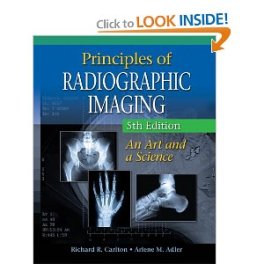
Principles of Radiographic Imaging: An Art and A Science 5e


Principles of Radiographic Imaging: An Art and A Science 5th Edition by Richard R. Carlton and Arlene McKenna Adler delivers essential info on radiographic contrast, density, detail, and distortion, as well as the newest instrumentation and know-how used within the imaging sciences. Constructing logically from the simplest ideas to the extra advanced, the book ties matters collectively visually and conceptually in a radical presentation of radiographic exposure.

Written in a student-friendly style, the text delivers content material logically, beginning with the best matters and steadily introducing extra advanced areas for study. Properly-recognized and revered in the field of radiologic sciences, authors Carlton and Adler share instructing and A.E.R.S. leadership expertise all through the text.

This book addresses each area of radiographic physics and instrumentation required by accredited programs in radiography, radiologist assistant, ultrasound, radiation remedy, and nuclear medicine, together with advanced areas of examine, including magnetic resonance imaging, computed tomography, bone densitometry, cardiac interventional, and other radiologic and imaging sciences.

The new chapters appear in Unit 6 covering the matters of Bone Densitometry, Nuclear Medicine, Radiation Remedy, and Diagnostic Medical Sonography. Discussions on recent developments in digital radiographic imaging and a whole chapter on radiology informatics methods give students the present data they have to be aggressive in at the moment's job market.

Customizable ancillaries embrace test banks, PowerPoint slides that coordinate with the text and its illustrations to make course preparation easier and more effective, and on-line course materials akin to CourseMate that embrace an interactive eBook, interactive teaching and learning tools such as flashcards and quizzes, and Engagement Tracker - a primary-of-its-form tool that displays student engagement in the course.


Download Principles of Radiographic Imaging PDF Ebook :

Senin, 17 November 2014

Comprehensive Radiographic Pathology 5th Edition, Eisenberg


Comprehensive Radiographic Pathology 5th Edition by Ronald L. Eisenberg and Nancy M. Johnson provides the essential pathology knowledge needed to produce quality radiographic images. It includes a general overview of physiology, and covers disorders and injuries by body system with descriptions of the pathology, its radiographic appearance, and treatment.

Now in full color, this reference includes the latest information on CT, MRI, SPECT, PET, ultrasound, and nuclear medicine, plus the always-popular Radiographer Notes with tips for digital imaging and patient care. The authors provide complete preparation for the classroom and for clinical practice! Comprehensive coverage helps you produce optimal images by understanding disease processes and their radiographic appearance.

High-quality radiographs and images clearly demonstrate the clinical manifestations of different disease processes and represent the quality images needed in radiography practice. A body systems approach makes it easy to locate and study information, with each chapter describing general physiology and then explaining various pathologic conditions and their radiographic appearance and treatment.

A discussion of specialized imaging techniques includes the full range of imaging modalities that may be needed to ensure correct diagnosis of pathologies, including ultrasound, computed tomography, magnetic resonance imaging, nuclear medicine, single-photon emission computed tomography (SPECT), or positron emission tomography (PET).

Treatment sections explain the most likely treatment for pathology along with its prognosis. Radiographer Notes include tips for producing optimal radiographs along with patient management recommendations. Summary Findings tables offer a quick reference to related pathologies, including location in the body, radiographic appearances, and treatments.

Study aids include an outline, key terms, objectives, and review questions for every chapter. Useful appendices include an extensive glossary, list of major prefixes, roots, and suffixes with definitions and examples. Updated content prepares you for today's practice with the latest on morbidity, mortality, and epidemiology; invasive squamous cell carcinoma; scoliosis; joint replacements; chest tubes and lines; MRSA (antibiotic-resistant staph infection); systemic and highly infectious diseases; more on fusion imaging, CT, MRI, SPECT, PET and correlation with radiography; new CT/MR images for correlation with radiographs; and Radiographer Notes updated with digital imaging information.

New full-color illustrations are added, and updated images add realistic detail for MRI, CT, sonography, and fusion PET/CT. Radiographic Pathology course enhances your understanding with interactive learning including animations, video clips, and narrated slide shows.


Download Comprehensive Radiographic Pathology PDF Ebook :

Radiation Protection in Medical Radiography 6th Edition


Radiation Protection in Medical Radiography 6th Edition by Mary Alice Statkiewicz Sherer, Paula J. Visconti and E. Russell Ritenour gives important info on radiation protection and biology in a transparent, concise, and straightforward-to-understand manner. Building from basic to more complex ideas, this book also presents radiation physics, cell construction, effects of radiation on people at the cellular and systemic levels, regulatory and advisory limits for human exposure to radiation, and the implementation of patient and personnel radiation safety practices.

Historic perspectives clarify the results of low-degree ionizing radiation and exhibit the link between radiation and cancer and different diseases. Chapter outlines and targets, highlighted key terms bulleted summaries, and review questions assist you to comply with and understand the material. Full-shade text and artwork programs improve visual appeal, reinforce vital elements, and hold your interest.

Evaluation questions with solutions allow you to assess your comprehension. Scholar Workbook helps you evaluate necessary text info introduced within the book. Companion online merchandise give you an internet supplement for the Sherer text. Updated NCRP and ICRP regulations provide the regulatory perspective you need for practice.

Discussion of Whole Efficient Dose Equivalent (TEDE) covers the radiation dosimetry quantity defined by the U.S. Nuclear Regulatory Fee to observe and control human publicity to ionizing radiation. Coverage of the Fukushima Daiichi Nuclear Plant Crisis addresses the influence of radiation ranges following Japan�s earthquake/tsunami in March 2011.

TRACE section covers the Instruments for Radiation Consciousness and Neighborhood Training program, a two-section approach to radiation dose consciousness and overall patient dose reduction via a three way partnership of AHRA and Toshiba�s Putting Patients First. Dialogue of the FDA white paper: Initiative to Cut back Unnecessary Exposure from Medical Imaging promotes the protected use of medical imaging devices, supports knowledgeable scientific choice making, and leads to elevated patient awareness.

This easy-to-learn text presents essential data on radiation protection and the biological results of ionizing radiation, to make sure its secure medical use. Constructing from basic to more complicated ideas, this book also presents radiation physics, cell structure, results of radiation on people at the cellular and systemic levels, regulatory and advisory limits for human exposure to radiation, and the implementation of patient and personnel radiation safety practices.


Download Radiation Protection in Medical Radiography PDF Ebook :

Merrill's Pocket Guide to Radiography 12th Edition


Merrill's Pocket Guide to Radiography 12th Edition by Eugene D. Frank, Barbara J. Smith and Bruce W. Long summarizes essential information for one hundred seventy of the most ceaselessly requested projections you'll encounter. Authors concisely provide just the data you will want for fast reference, preserve it with you and keep Merrill's shut at hand!

Diagnostic-high quality radiographs demonstrate desired imaging results. Key positioning data is formatted for quick and simple access. Each procedure is presented in a two-color, two-page spread with bulleted, step-by-step procedures and accompanying photographs on the top page; and a chart with spaces to fill within the particular methods used for a specific projection on the bottom page.

Section dividers with tabs supply fast access to every section. Computed radiography information allows you to make the delicate adjustments essential to obtain optimal outcomes with CR. Publicity technique chart for every projection helps cut back the variety of repeat radiographs and improves overall picture quality. Abbreviations and external landmark charts on the within covers present quick entry to incessantly needed information.

The kVp values are included for each projection. Compensating filter info is included for those projections the place filters are used. There are new publicity index column for use with digital imaging programs and particular collimation settings for all projections executed using DR Systems. This pocket guide is designed to be used in the clinic to help technologists effectively and precisely produce quality diagnostic images.

The book was written for radiology college students and technologists. It is a quick and handy useful resource for positioning on all kinds of radiologic procedures. This edition has added a section on obese patients, which is helpful for our numerous patients. This can be useful and much-wanted information for radiology technologists and students.


Download Merrill's Pocket Guide to Radiography PDF Ebook :